Oral pathology in Denver, CO
Colorado Oral Surgery provides evaluation and diagnosis for oral pathology in Denver, CO. Oral pathology focuses on identifying diseases of the mouth, jaws, and related structures, including the lips, tongue, salivary glands, and facial bones.
About oral pathology evaluation
Oral pathology examines the cause and nature of conditions that affect the mouth and jaws. Common concerns include non-healing ulcers, red or white patches, lumps, pigmented spots, bite changes, or unexplained pain.
Many lesions are harmless, but some can represent precancer or cancer. Early detection is important for timely care and better outcomes.
How oral pathology can help you
Clarifies the nature of a mouth lesion through clinical exam and testing
Supports early detection of oral cancer and precancerous changes
Guides the right treatment plan, from monitoring to surgery or referral
Reduces uncertainty with a clear diagnosis and evidence-based next steps
What to expect
Before a biopsy, you may be asked to pause certain blood thinners if medically safe and to eat a light meal. During the procedure, local anesthetic numbs the area. After, expect minor soreness for a day or two.
See a clinician promptly if a mouth sore lasts longer than two weeks, bleeds easily, or keeps returning.
Signs that merit an oral pathology visit
- Ulcer or sore that does not heal within 14 days
- Red, white, or mixed red-white patches (erythroplakia or leukoplakia)
- Lump, thickened tissue, or rough area on the tongue, cheek, or gums
- Unexplained numbness, pain, or loosening of teeth
- Jaw swelling, persistent hoarseness, or difficulty swallowing
The oral pathology assessment process
1) Medical and dental history
Reviewing medications, tobacco and alcohol use, systemic conditions, and prior biopsies.
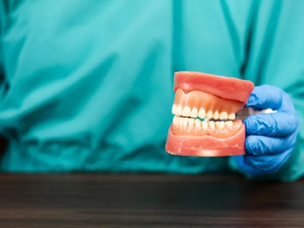
2) Head and neck exam
Checking lips, cheeks, tongue, floor of mouth, palate, gums, salivary glands, and lymph nodes.
3) Imaging and documentation
Dental x-rays or 3D scans to assess bone, teeth roots, or cyst-like changes. Photographs track lesion appearance over time.
4) Biopsy and pathology report
A small tissue sample is removed under local anesthesia for microscopic analysis. Results are typically available within several days to two weeks.