Digital X-rays in Denver, CO: a patient guide

Colorado Oral Surgery uses digital X-rays to support accurate diagnosis and safe treatment planning for patients in Denver, CO, and Parker, CO. Digital X-rays provide detailed images with a low radiation dose and quick viewing.
Digital dental imaging explained
Digital X-rays, also called digital dental radiography, use an electronic sensor instead of film to capture images of your teeth, bone, and jaw joints. The sensor converts X-ray energy into a digital image that appears on a computer within seconds.
Compared with traditional film, digital X-rays typically use significantly less radiation while producing high-resolution images.
Why consider digital X-rays?
Lower radiation dose than conventional film
Fast results with images available in seconds
Clear detail to spot decay, infection, and bone changes early
Easy image sharing for referrals or second opinions
Your imaging experience
Most patients find digital X-rays quick and comfortable. Bitewings and periapicals involve briefly holding a sensor with gentle guidance. A panoramic unit rotates around your head without placing anything inside the mouth.
Safety and radiation basics
- Digital systems keep exposure as low as reasonably achievable (ALARA principle)
- Modern sensors are sensitive, reducing the amount of radiation required
- Protective aprons and thyroid collars are used when appropriate
- Pregnancy considerations are discussed to time imaging safely
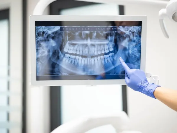
Images are available immediately with a clear explanation of findings and how they relate to your treatment plan.
The digital X-ray process
1) Preparation
Your visit starts with protective measures such as a lead apron and, when appropriate, a thyroid collar.
2) Sensor placement
A small sensor is placed in the mouth or positioned outside the mouth, depending on the image needed (bitewing, periapical, panoramic, or CBCT).
3) Image capture
Each exposure takes only a moment. The digital image appears on the computer within seconds.
4) Review
The team reviews the images with you, pointing out findings such as decay, bone loss, or impacted teeth.